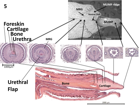
FIG. 5.
Scanning electron micrograph (top), transverse H&E sections (middle) and midsagittal H&E section (bottom) of the glans penis. Arrows indicate exact corresponding location between transverse and longitudinal sections. Modified from Yang et al. 2010 [8].

