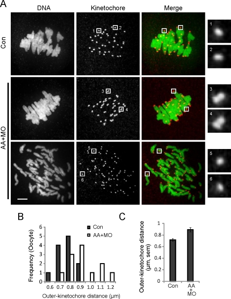
FIG. 2.
Centromeres are more susceptible than chromosome arms to cohesin loss. A) Control oocytes (Con; n = 12) or oocytes microinjected with both AA-separase cRNA and securin MO (AA+MO; n = 23) were fixed 8 h after meiotic resumption and stained for DNA (green) and kinetochores (CREST; red). The AA+MO group includes oocytes with intact chromosomes (middle; n = 12 of 23) and prematurely separated chromosomes (bottom; n = 11 of 23). Images are maximal intensity projections of confocal z-series, with insets of single optical sections to show kinetochores. Insets 1–4 include the two kinetochores of a sister pair; insets 5 and 6 show single kinetochores in AA+MO oocytes where chromosomes prematurely separated. Oocytes were collected from a total of four mice. Bar = 5 μm. B and C) Outer kinetochore distances were measured from the outer edges of sister kinetochore pairs for control and AA+MO oocytes with intact chromosomes (n = 240 kinetochore pairs from 12 oocytes in each group). The populations are represented by histograms (B) and by the mean ± SEM for each group (C).

