FIG. 3.
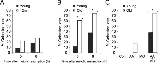
Cohesion is reduced in old oocytes. A) Young (n = 22 from four mice) and 12m oocytes (n = 18 from eight mice) microinjected with a cRNA-encoding fluorescently labeled H2B, AA-separase cRNA, and securin MO to increase separase activity were matured in vitro and imaged live by fluorescence at 5 and 8 h after meiotic resumption. Percentage of oocytes with cohesion loss, as indicated by prematurely separated bivalents and chromatids, was determined for each group. At both time points, bivalents in young and 12m oocytes are similarly intact. B) Young and old oocytes were microinjected as in A and imaged at 5 h (n = 17 young and 13 old) and 8 h (n = 29 young and 20 old) after meiotic resumption. Percentage of oocytes with cohesion loss is significantly higher in old oocytes compared to young (asterisks) at both 5 h (Fisher exact test, P = 0.007) and 8 h (P = 0.019). Oocytes were collected from 11 young and 10 old mice. C) Young and old oocytes were microinjected with a cRNA-encoding fluorescently labeled H2B (Control; n = 94 young and 14 old), AA-separase cRNA (AA; n = 6 young and 12 old), securin MO (MO; n = 13 young and 9 old), or both AA and MO (AA+MO; n = 29 young and 20 old) and imaged at 8 h after meiotic resumption. The increased cohesion loss in old AA oocytes is not statistically significant. Oocytes were collected from 34 young and 34 old mice.

