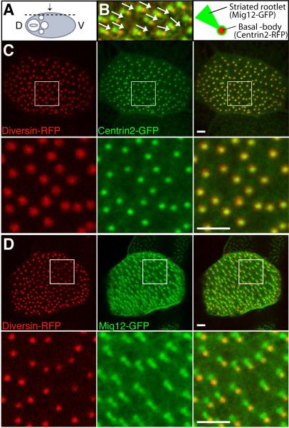
Fig. 1. Diversin localizes to the basal body in multi-ciliated cells.
Both ventral blastomeres of four-cell embryos were injected with the following mRNAs as indicated: Div-RFP (0.1–0.2 ng), Centrin2-RFP (0.4 ng), Centrin2-GFP (0.1 ng) or Mig12-GFP (0.1 ng). Injected embryos were fixed at stage 34 and cryosectioned for confocal imaging. (A) Experimental scheme. Plane of sectioning is indicated by a dashed line; arrow shows direction of viewing. D, Dorsal; V, Ventral. (B) Mig12-GFP and Centrin2-RFP label the striated rootlet and the basal body, respectively, as indicated in the cartoon on the right. Arrows represent basal body polarity. (C, D) Div-RFP localizes to the basal bodies labeled with Centrin2-GFP (C), but not to the striated rootlets marked by Mig12-GFP (D). Lower panels show boxed images at higher magnification, merged files are on the right (B–D). Representative confocal images of embryonic epidermis are shown. Scale bar, 2 μm.

