
Fig. 3. Diversin is required for basal body polarity and ciliogenesis.
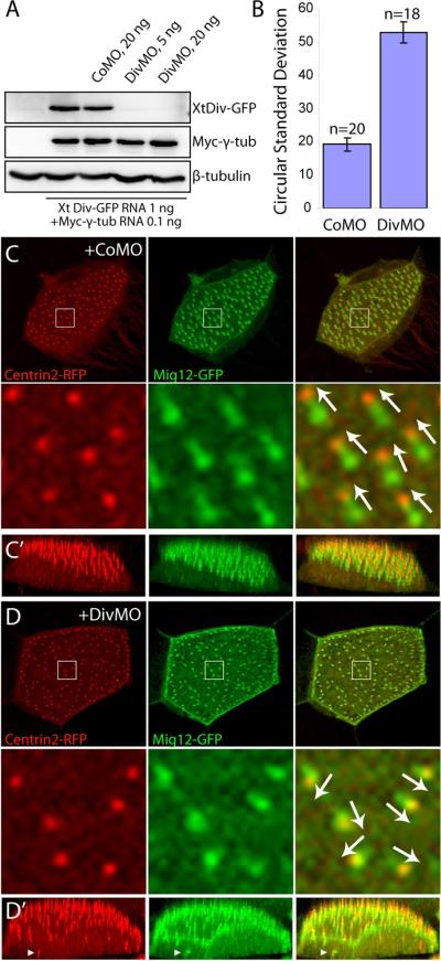
(A) Specificity of DivMO. Embryos were injected with CoMO or DivMO, as indicated, together with XtDiv-GFP RNA (1 ng) and myc-γ tubulin RNA (0.1 ng). Western analysis of st. 11.5 embryo lysates shows specific inhibition of XtDiv-GFP by DivMO, but not CoMO. XtDiv-GFP was detected with anti-GFP, whereas myc-γ tubulin was detected with anti-Myc antibody. Anti-β-tubulin antibody was used to control loading. (B–D) MOs (20 ng) and RNAs encoding Centrin2-RFP (0.4 ng) and Mig12-GFP (0.1 ng) were injected into the animal-ventral region of four-cell embryos. (B) Basal body polarity was quantified in a representative experiment by calculating circular standard deviations (see Materials and Methods). Results are shown as means +/− SEM (n = 20 and 18 for CoMO and DivMO respectively). (C, D) The apical regions of multi-ciliated cells scored in (B) are shown as x–y (C, D) or x–z plane (C', D') projections of serial optical sections (see also Fig. 1 legend). Arrows indicate basal body polarity. Top is apical in C' and D'. (C, C') CoMO does not affect basal body apical localization and polarity. (D, D') DivMO disrupted basal body polarity and striated rootlet structure. Arrowheads point to defects in basal body apical docking.