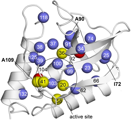
Fig. 1.
Structure of Δ+PHS nuclease showing internal positions that were substituted with Arg. Spheres represent the Cα of residues substituted with Arg. Colors identify variants that were unfolded by the substitution with Arg (red), variants that were folded but enzymatically inactive (yellow), and variants that were folded and enzymatically active (blue). Bold numbers identify the positions (72, 90, and 109) that were substituted with Arg in the three crystal structures that were solved. The circle identifies the approximate location of the active site. Structure shown is Δ+PHS (PDB ID code 3BDC) (42).

