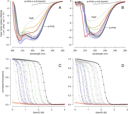
Fig. 2.
Far-UV CD spectra of Arg-containing variants at pH 7 (A) and pH 10 (B). Δ+PHS nuclease (solid black line), Δ+PHS nuclease in 6 M GdnHCl (dashed black line), and the unfolded SNase variant T62P in water (dashed gray line) are shown for reference. Solid gray lines correspond to the 19 Arg variants with no apparent structural rearrangement. Variants of interest are highlighted: A58R (green); V39R, Y91R, and N100R (blue); I92R (orange); and V104R (red). GdnHCl-induced unfolding of Arg-containing variants at pH 7 (C) and pH 10 (D). Points represent the normalized fluorescence measured at 296 nm; lines represent two-state linear extrapolation model fit to each curve. Color scheme is identical to A and B. The two-state model could not be fit to the titration curves of the I92R or V104R variants.

