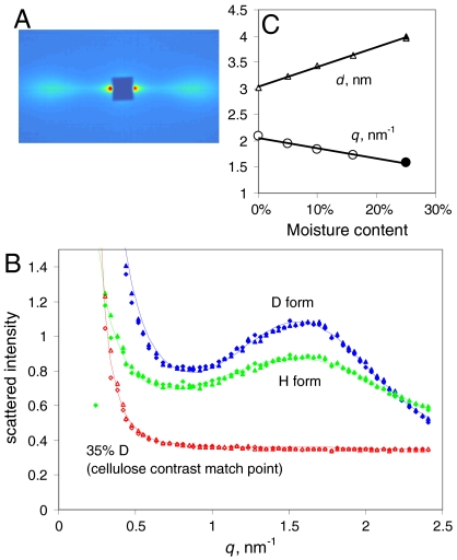
Fig. 1.
SANS from spruce wood. (A). SANS pattern at 25% hydration with D2O, showing equatorial Bragg reflections at q = 1.6 nm-1. The fiber axis is vertical. (B). Radial profiles of SANS intensity at 25% hydration with D2O, H2O and a 35∶65 mixture of D2O with H2O, matching the scattering length density of cellulose based on its elemental composition. (C). Variation in position of the center of the fitted radial intensity peak with the level of hydration with D2O, and the corresponding d-spacings between microfibril centers.

