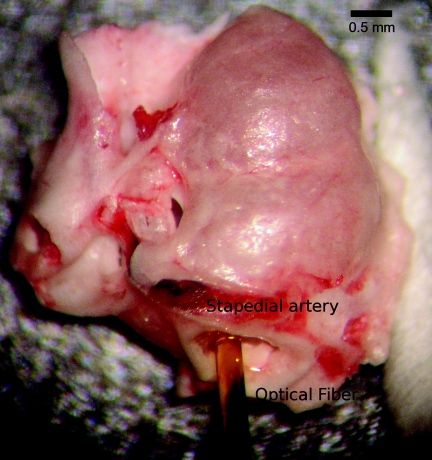
Figure 1.
Placement of the optical fiber for tone-on-light masking experiments. An ex vivo image of the 200-μm diameter optical fiber inserted into the gerbil cochlea. The optical fiber was abutted to the round window membrane, but did not puncture it. The orientation was toward the lateral position of the round window (left in this picture). Various tuning curves were acquired with the optical fiber in different positions in the round window along the trajectory from lateral to medial (left to right). The fiber was mounted on an x-y-z translator coupled with a micromanipulator. (See also Ref. 18 for optical fiber placement.)

