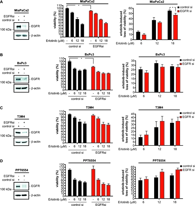
Figure 2.
Erlotinib response of PDAC cells is EGFR independent. MiaPaCa2 (A), BxPc3 (B), T3M4 (C), and PPT6554 (D) cells were transfected with a control siRNA or an EGFR-specific siRNA. Left panel: Knockdown of the EGFR 48 hours after the transfection by Western blot analysis. β-Actin controls equal protein loading. Twenty-four hours after transfection, cells were treated with increasing doses of erlotinib as indicated for 48 hours or were left as an untreated control. Viability was determined using MTT assays. Middle panel: Viability of control siRNA-transfected cells was arbitrarily set to 100% and compared to (I) control siRNA-transfected and erlotinib-treated cells, to (II) EGFR siRNA-transfected cells, and to (III) EGFR siRNA-transfected and erlotinib-treated cells. Right panel: Erlotinib-induced therapeutic response in control and EGFR siRNA-transfected cells. Note that the right panels rely on an extrapolation of the data presented in the middle panels to better visualize the erlotinib-induced loss of viability (=therapeutic response) in control and EGFR siRNA-transfected cells. Student's t test, *P < .05 versus controls.

