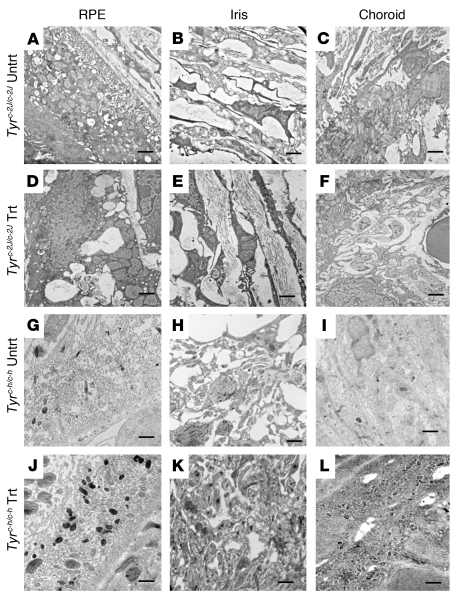
Figure 5. TEM images of RPE, iris, and choroid of treated Tyrc-2J/c-2J and Tyrc-h/c-h mice.
Tyrc-2J/c-2J mice showed no discernible pigment in melanosomes, either in the absence of treatment (A–C) or after 1 month of nitisinone treatment (D–F). In contrast, Tyrc-h/c-h mice showed an appreciable increase in the number of pigmented melanosomes after 1 month of nitisinone treatment (J–L) compared with untreated mice (G–I). Scale bars: 500 nm. Original magnification, ×12,000.

