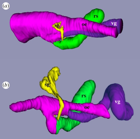
Figure 2.
Three dimensional reconstructions of transverse sections through the foregut of (a) a late-stage larva of Conus lividus and (b) a post-larva at 48 h after the onset of metamorphosis showing the separation of the venom gland (vg) from the mid-oesophagus (oe). Anterior is towards the left for both. The posterior extent of the reconstructions ends where the mid-oesophagus began to coil around the columella of the shell. Other abbreviations: rs, radular sac; sg, salivary glands.

