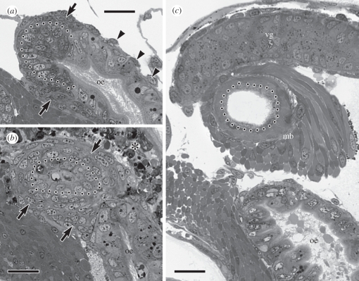
Figure 4.
Development of the muscular bulb of the venom apparatus; dotted line in all images demarcates the basal epithelial surface of the venom gland. (a) Oblique section through the mid-oesophagus (oe) as it begins to coil around the columella showing accumulation of myoblasts (arrows) around the posterior extremity of the future venom gland; arrowheads indicate nuclei of sparse oesophageal muscle cells. (b) Section through the same area of the mid-oesophagus at 24 h after velum loss showing recent separation of the venom gland from the mid-oesophagus and multi-layered myoblast sheath (arrows) around the terminus of the venom gland. The asterisk indicates degenerating cells of the velar lobes that have involuted into the head. (c) Section through the same area of the mid-oesophagus at 4 days after velum loss showing a secretory portion of the venom gland (vg) and the fully differentiated muscular bulb (mb). Scale bars: (a–c) 20 µm.

