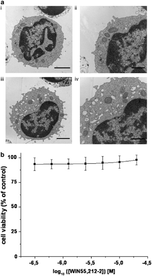
Figure 7.
WIN55,212-2 induced vacuolation in PB1 cells. (a) Normal ultrastructure morphology of primary PB1 cells was predominately found in cells treated with the vehicle for 24 h. These cells had a well-defined plasma membrane and uniformly distributed chromatin in the nucleus and well-defined organelles in the cytoplasm (i and ii). Cells treated with WIN55,212-2 for 24 h displayed vacuoles in the cytoplasm. Bars; i and iii=2 μm; ii and iv=1 μm. (b) PB1 cell viability (XTT) in response to WIN55,212-2 for 48 h. The experiment was performed once in triplicates

