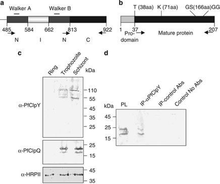
Figure 1.
PfClpQ and PfClpY are expressed in the asexual-stage parasites and form a protein complex in the parasite. (a) Schematic domain structure of PfClpY (Gene ID PFI0355c) showing location of N, I and C domains, respective amino-acid positions and walker A and B domains are also indicated. (b) Schematic representation of structure of PfClpQ showing location of pro-domain and mature protease. The locations of conserved residues of active site triad are also marked. (c) Western blot analyses of total lysates of equal number of synchronized parasites at ring (8–10 h post invasion (hpi)), trophozoite (30–32 hpi) and schizont (42–45 hpi) stages with antibodies against the PfClpQ and PfClpY showing expression of both the proteins in trophozoite-schizont stages. Anti-histidine rich protein-II (HRPII) antibodies were used to probe a blot run in parallel to show equal loading in each well. (d) Co-immunoprecipitation of PfClpQ and PfClpY from parasite lysate using anti-PfClpY antibodies; PfClpQ protein was detected in the immunoprecipitated protein complex by western blot analysis. Immunoprecipitation reactions using non-specific antibodies or no antibodies were used as controls

