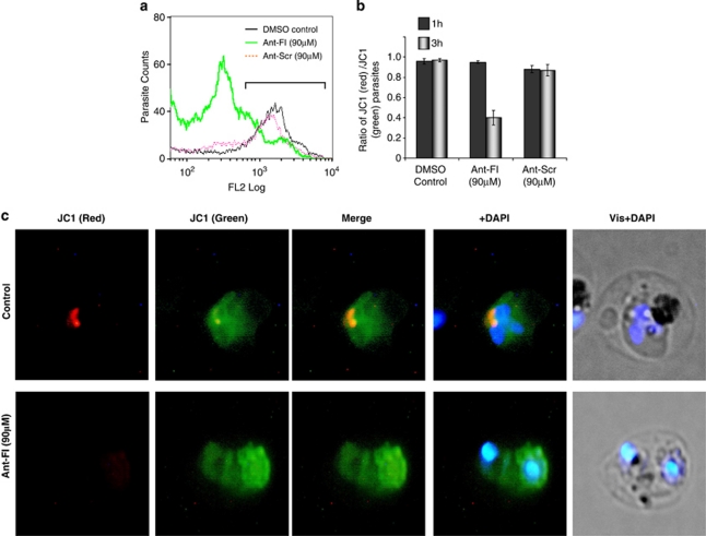
Figure 4.
In situ mitochondrial membrane potential of P. falciparum parasite after treatment with Ant-FI-peptide as estimated by JC-1 staining. (a) Flow-cytometry histogram showing reduction in JC-1 red staining in the parasite population treated with Ant-FI-peptide as compared with parasites treated with scrambled peptide or solvent alone. (b) Graph showing ratio of JC-1 (red)/JC-1 (green) parasite population after treatment with Ant-FI, Ant-Scrambled (Ant-Scr) or solvent alone. (c) Fluorescent microscopic images of JC-1-stained parasite showing accumulation of aggregated JC-1 (red staining) in the mitochondria and monomeric JC-1 (green staining) in the cytosol. Treatment of Ant-FI-peptide caused loss of membrane potential, which reduced red staining in the mitochondria of the parasite. The parasite nuclei were stained with DAPI (blue) and parasites were visualized by fluorescence microscope

