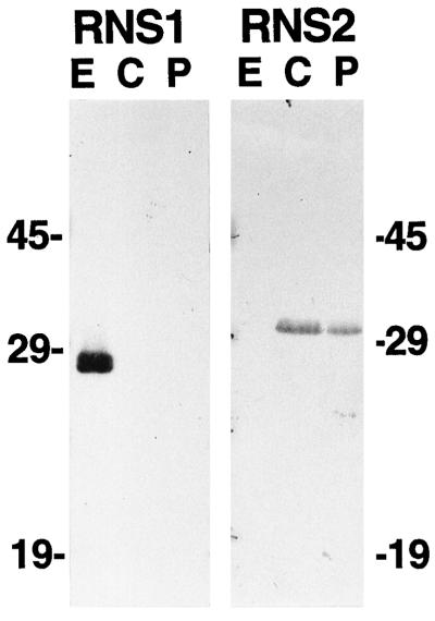
Figure 4.
Extra- and intracellular localization of RNS1 and RNS2. Protein samples were prepared from Arabidopsis cell cultures as described in Methods. Seventy-five micrograms of protein per lane was separated on SDS-PAGE gels, transferred to a membrane, and immunodetected with anti-RNS1 or anti-RNS2 serum. E, Extracellular fraction; C, whole-cell lysate; P, protoplast lysate.

