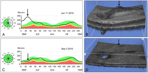
Fig. 3.
(A) An optical coherence tomography retinal nerve fiber layer scan in the left eye demonstrated retinal nerve fiber layer (RNFL) thickening of the superotemporal region (arrow) rather than RNFL thinning. (B) Optical coherence tomography (OCT) 3-D reconstruction imaging shows superotemporal peripapillary retinoschisis as a 3-D (arrow). (C) After 3 months of topical prostaglandin analogue medication, an OCT RNFL scan revealed a decrease in RNFL (arrow). (D) OCT 3-D reconstruction imaging shows the decreased peripapillary retinoschisis (arrow). TEMP=temporal; SUP = superior; NAS = nasal; INF = inferior.

