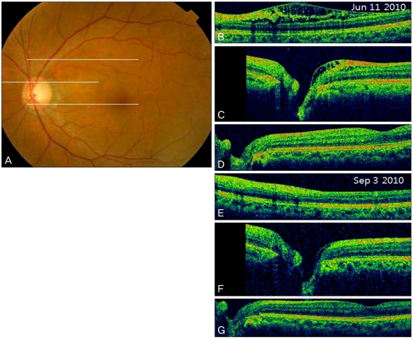
Fig. 4.
(A) Fundus photography. (B) Optical coherence tomography (OCT) slice at the superotemporal region from the optic disc (upper white line in A) shows inner retinal schisis cavities. (C) OCT slice at the level of the superior part of the optic disc and some part of the macula (middle white line in A) shows the peripapillary retinoschisis, not extending to the macula. (D) OCT slice at the level of the inferior part of the optic disc and the fovea (lower white line in A) shows that there is no retinoschisis in the papillomacular retinal region. (E,F,G) After 3 months of topical prostaglandin analogue medication, the OCT scan shows that the peripapillary retinoschisis is partially resolved.

