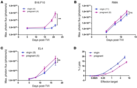
Figure 6. NK cell impairment underlies increased metastasis in pregnant immunocompetent C57BL/6 mice.
(A–C) Bioluminescence imaging after tail vein injection of 5 × 10⁵ luciferase-expressing syngeneic, NK-sensitive B16.F10 melanoma cells (A) and NK-resistant RMA (B) and EL4 (C) lymphoma cells into 14-day pregnant and virgin C57BL/6 mice. **P < 0.01. (D) Standard 4-hour 51Cr release cytotoxicity assay with splenocytes extracted from poly(I:C)-stimulated 16-day pregnant and virgin C57BL/6 mice and Yac-1 target cells. Graph was adjusted for NK proportions in splenocytes, as determined by flow cytometry. 4 mice were used per condition.

