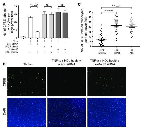
Figure 4. Role of eNOS for the effects of HDL on TNF-α–stimulated endothelial monocyte adhesion.
(A) Inhibition of eNOS-mediated NO production by siRNA-specific knockdown of eNOS or treatment with L-NAME (1 mM) prevented the potent inhibitory effects of HDL from healthy subjects on endothelial cell monocyte adhesion (n = 6–8 per group). Endothelial cells were incubated with HDL (50 μg/ml) from healthy subjects, and binding of CFSE-labeled human monocytes was analyzed after 3 hours by fluorescence microscopy (HAECs were stimulated with TNF-α; 5 ng/ml). (B) Representative photographs (original magnification, ×10) of endothelial monocyte adhesion obtained by fluorescence microscopy are shown. (C) Effects of HDL isolated from patients with sCAD and ACS as compared with HDL from healthy subjects on TNF-α–stimulated endothelial monocyte adhesion (n = 25 per group; data points for all study participants are shown).

