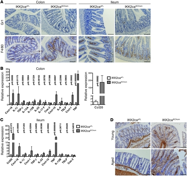
Figure 2. Immune cell infiltration and increased cytokine and chemokine expression in the gut of IKK2caIEChom mice.
(A) Immunohistochemical staining for Gr1 and F4/80 reveals increased numbers of granulocytes and macrophages, respectively, in colonic and SI cross sections of 6-week-old IKK2caIEChom mice. In the SI of IKK2caIEChom mice, F4/80-positive cells accumulated around the crypts, while in control mice, macrophages were mainly found in the villi. (B and C) qRT-PCR analysis shows enhanced expression of a subset of proinflammatory genes in the colon (B) and the ileum (C) of 7- to 8-week-old IKK2caIEChom mice (n ≥ 6 for each genotype; mRNA levels are presented as mean ± SD). (D) Immunohistochemical staining with antibodies recognizing α-SMA revealed the presence of increased numbers of activated myofibroblasts in colons of young (10 week old) and aged (1 year old) IKK2caIEChom mice compared with IKK2casFL littermates. Scale bars: 50 μm.

