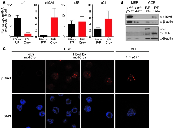
Figure 5. Loss of the Lrf gene leads to p19Arf upregulation in GCB cells.
(A) GCB cells were FACS sorted and mRNA levels of Lrf, p19Arf, p53, and p21 measured by quantitative RT-PCR and normalized to corresponding Hprt1 mRNA levels. Data represent mean with SD. (B) GCB cells were FACS sorted and Western blot for p19Arf, Lrf, Irf4, and β-actin performed. Protein extracts obtained from Lrf–/–p53–/– and Lrf–/–p19Arf–/– MEFs were used as positive and negative controls for p19Arf expression, respectively. Irf4 protein was only detected in GCB cell samples, confirming enrichment of GCB cells. Lrf protein was only detected in control GCB cells. (C) Immunofluorescence analysis of p19Arf protein in FACS-sorted GCB cells. p19ARF protein accumulated in nucleoli of Lrf-deficient GCB cells. Lrf–/–p53–/– MEFs are shown as a positive control for p19Arf stain (red). Nuclei were counterstained with DAPI (blue). Original magnification, ×630.

