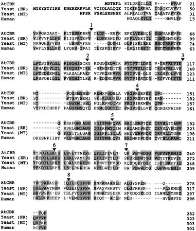
Figure 1.
The multiple alignment of amino acid sequences of AtCBR with those of NADH-Cyt b5 reductase from human (Yubisui et al., 1984) and ER (Csukai et al., 1994) and mitochondria (MT) (Hahne et al., 1994) of yeast. Shading indicates the conserved amino acid residues among the aligned sequences. The arrowheads and numbers over the AtCBR sequence indicate positions where introns are inserted in the AtCBR gene. The sequence of the AtCBR cDNA and the AtCBR gene were deposited in the databank under accession nos. AB007799 and AB007800, respectively.

