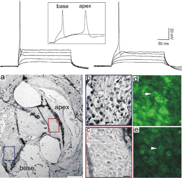
Fig 2.
Endogenous electrophysiological firing patterns and voltage-dependent ion channel composition differ between apical and basal spiral ganglion neurons. Top Panel. Series of stacked whole-cell current clamp traces from a basal (left) and apical (right) spiral ganglion neuron highlight differences in response speed. The onset time course and difference in latency are evident from the series of sweeps; the differences in action potential duration can be observed from the inset traces (box). a–e, Anti-BK antibody labeling was enriched in basal compared to apical spiral ganglion neurons in adult cochlea (a–c) and in vitro (d, e). a, Section taken from an adult CBA/CaJ mouse cochlea stained with anti-BK antibody (Alomone Labs, APC-02) showing that the neurons in the base of the cochlea (blue box) were considerably darker than those in the apex (red box). The calibration bar, lower right, represents 200 μm. b, high-magnification view of the basal neurons. c, High-magnification view of the apical neurons. d, postnatal spiral ganglion neurons isolated from the base and maintained in vitro for 7 days also showed intense anti-BK antibody labeling. The arrow indicates a darkly stained neuron. e, Sister cultures of apical neurons treated identically to those in panel d showed only low staining levels. The arrowhead indicates a lightly stained neuron. The scale bar in panel c = 20 μm and applies to panels b–e. Adapted from Adamson et al., 2002a.

