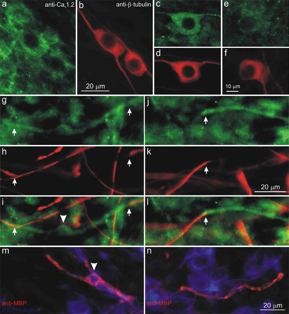
Figure 10.
The Cav2.3 α-subunit was present in both neurons and satellite cells in the spiral ganglion and was associated with compact myelin. (a) Anti-Cav2.3 antibody (green) stained for neurons and satellite cells. (b, d, f, h, k) Anti-β-tubulin antibody (red) was used as a neuron-specific marker. Instances in which surrounding satellite cells did not preclude visualization, anti-Cav2.3 antibody (green) was enriched in a small number of neurons (c), while most neurons (e) were lightly labeled. (g, j) anti-Cav2.3 immunofluorescence (green) was detected in a compact myelin-like structure (between arrows) that surrounded (h, k) anti-β-tubulin-labeled neurites. (i,l) Overlay images from g, h and j, k, respectively (arrowhead in i indicates the soma profile of a putative Schwann cell. (m, n) Overlaid images of anti-myelin basic protein (SMI-99, red) surrounded neuronal processes (anti-β-tubulin, blue), which appeared similar to structures labeled by anti-CaV2.3 in panels g–l. Arrowhead (m) indicates the soma profile of a myelinating Schwann cell. Calibration bar in b applies to a–b; f applies to c–f; k applies to g–l; n applies to m–n.

