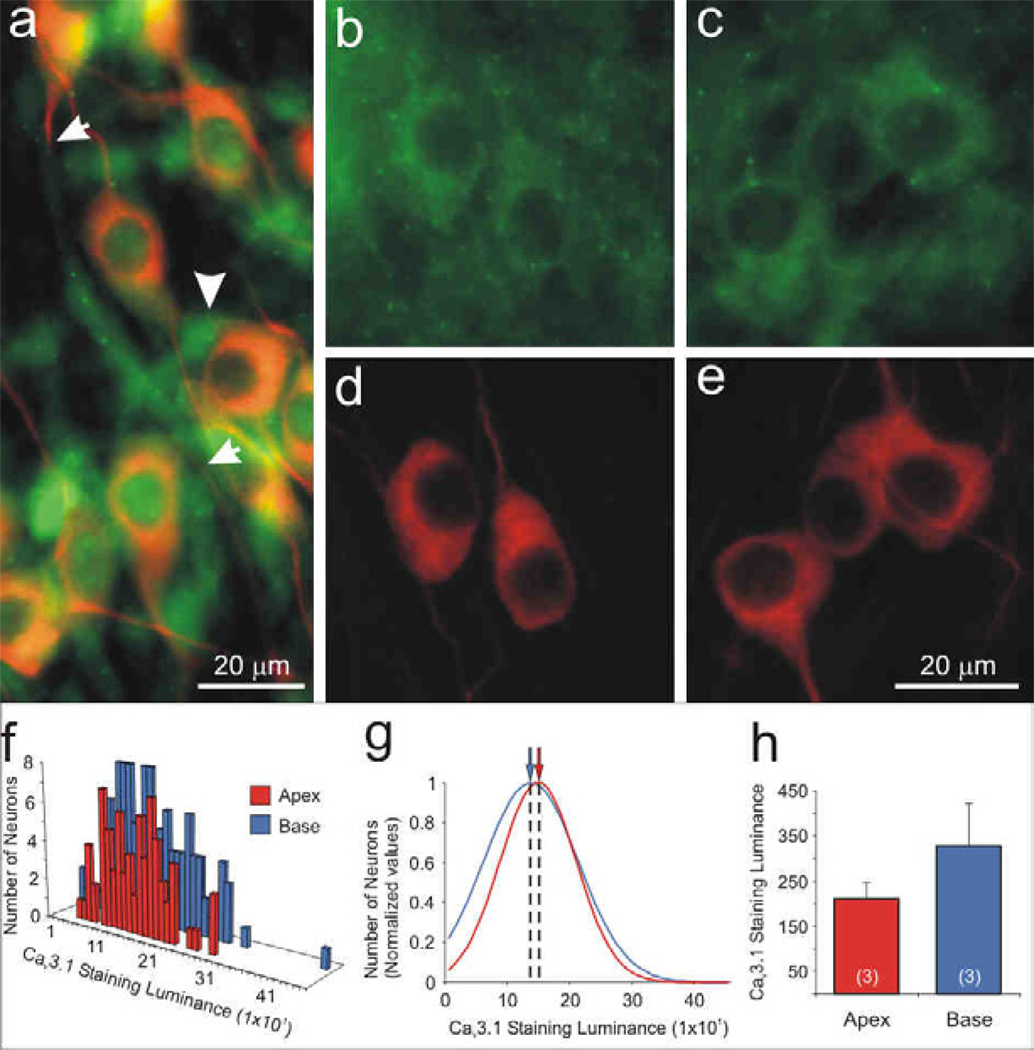
Figure 11.
The Cav3.1 α-subunit was localized to satellite cells, compact-myelin-like structures, and uniformly labeled apical and basal spiral ganglion neurons. (a) anti-Cav3.1 antibody labeled satellite cells diffusely (arrowhead) as well as regions detected in a compact myelin-like structure (between arrows) that surrounded an anti-β-tubulin-labeled neurite (red). In areas with lower levels of surrounding satellite cells, we were able to detect neuronal labeling with anti-Cav3.1 antibody (green), which showed similar intensity levels in (b) apex and (c) base neurons. (d–e) Anti-β-tubulin antibody (red). Anti-Cav3.1 antibody staining luminance is illustrated by (f) representative histograms and (j) normalized Gaussians for a single experiment. (h) Averaged anti-Cav3.1 immunofluorescence did not differ systematically between apical and basal neurons (n = 3; p > 0.20). Calibration bar in panel e applies to b–e.

