Figure 2.
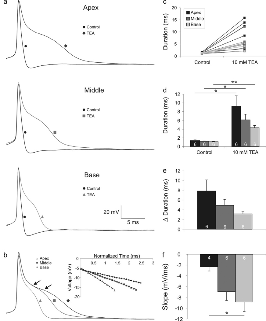
Action potential repolarization is prolonged differentially in apical, middle, and basal neurons after tetraethylammonium (TEA) application. (a) Representative recordings from apical, middle, and basal neurons before (black traces labeled with circles) and after application of TEA (gray traces labeled with a diamond, square and triangle, respectively). (b) Overlaid traces of apical, middle, and base whole-cell current clamp recordings exemplifying the differences in action potential repolarization. Arrows indicate the region of measurement for slope values. Inset: calculated slope of the plateau region; R2 > 0.99 for all fits. (c) Individual duration measurements before and after TEA application. (d) Averaged measurements show significant action potential duration increases in the apex, middle, and base after TEA application. (e) Change in action potential duration before and after TEA application shows that the relative action potential duration between the apex, middle and basal neurons is retained. (f) Averaged slope measurements show a significant difference between the apical and basal neurons. (For this and subsequent figures, paired Student’s t-test: *, p < 0.05; **, p < 0.01).

