Figure 5.
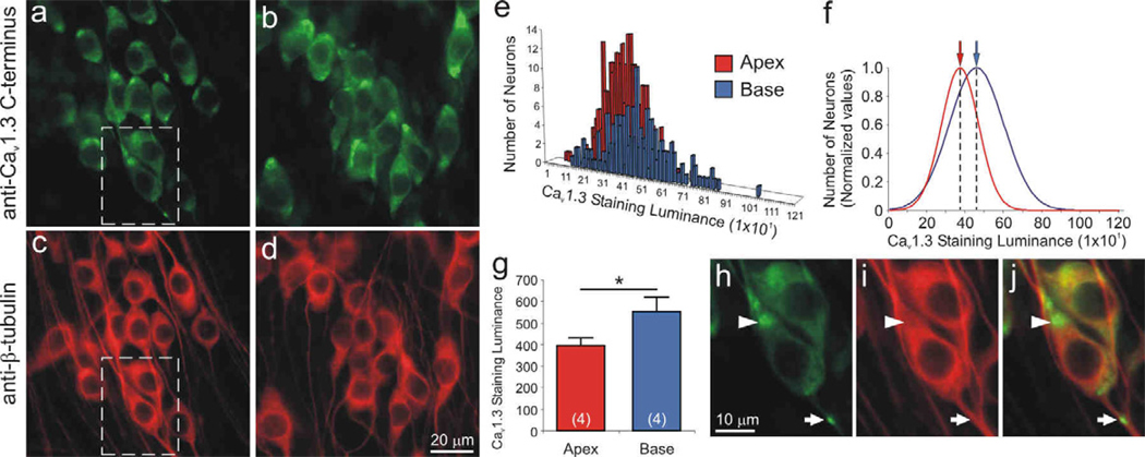
The Cav1.3 α-subunit was localized predominantly in basal spiral ganglion neurons. Neurons labeled with anti-Cav1.3 C-terminus antibody (green) showed differential immunofluorescence in (a) apex and (b) base neurons. (c, d, i) Anti-β-tubulin antibody (red) was used as a neuron-specific marker, which showed relatively uniform labeling. Dotted area in panels a and c was magnified in panel h and i, respectively. (e) Representative histograms of apex and base Cav1.3 staining luminance in a single experiment were (f) normalized and fitted with Gaussians; means for apical and basal neurons indicated by arrows (red and blue, respectively). (g) Averaged anti-Cav1.3 antibody luminance (n = 4) was significantly higher in the base than in the apex (*, p < 0.05). (h) Anti-Cav1.3 antibody was clustered at the poles of cell somata (arrowhead) and occasionally along neurites (arrow). (j) Overlay of β-tubulin and Cav1.3 staining luminance. Calibration bars in panels d and h apply to panels a–d and h–j, respectively.

