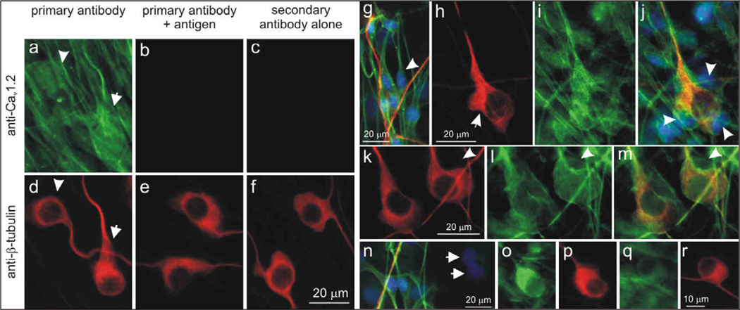
Figure 8.
The Cav1.2 α-subunit was present in both neurons and satellite cells in the spiral ganglion and was associated with myelination. (a) Control experiments confirmed the specificity of anti-Cav1.2 antibody. Neurons (arrow, arrowhead) and satellite cells were labeled with anti-Cav1.2 antibody (green). (b) Antibody pre-incubated with control antigen peptide showed little or no anti-Cav1.2 antibody labeling. (c) Little or no immunofluorescence was observed in secondary antibody alone controls. (d–f) Anti-β-tubulin antibody (red) was used as a neuronal marker. (g) Anti-CaV1.2 antibody (green) labeled the soma and processes of bipolar spindle-shaped satellite cells (arrowhead) with a morphology that resembled Schwann cells. Hoechst dye (blue) was utilized to highlight cell nuclei, neurons and their processes were labeled with anti-β-tubulin (red). (h–j) Schwann cells surrounded the cell body of a neuron (arrowhead) and were aligned along neuronal processes (h) anti-β-tubulin (red), (i) anti-CaV1.2 (green), (j) overlay with Hoechst dye (blue). (k–m) A sub-set of neurons were encapsulated by CaV1.2 immunofluorescent structures resembling loose myelin sheaths that surround spiral ganglia somata; arrowheads indicate a putative Schwann cell soma. (k) anti-β-tubulin (red), (l) anti-CaV1.2 (green), (m) overlay. (n) Two non-immunoreactive satellite cells (arrows); overlay of anti-Cav1.2 (green), anti-β-tubulin (red), Hoechst dye (blue). Neurons stained with different intensities: (o) brightly-stained neuron, (q) lightly-stained neuron. (p, r) Anti-β-tubulin antibody. Calibration bar in panel a applies to a–f; h applies to h–j; k applies to k–m; r applies to o–r.

