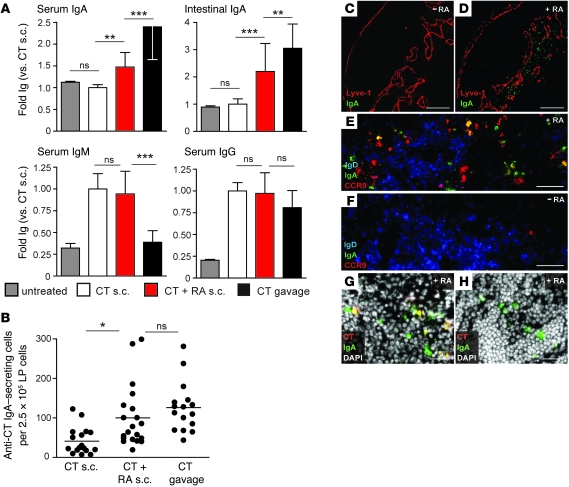
Figure 4. Skin-draining ingLNs of RA-treated mice support induction of gut-homing receptor and IgA expression on B cells.
Mice were immunized on days 0 and 10 either s.c. with 1 μg CT or orally with 10 μg CT. A group of s.c. immunized mice additionally received s.c. RA injections on days 0, 1, 2, 3, 6, 10, and 13 after antigen delivery. On day 14, mice were sacrificed. (A) Levels of CT-specific Ig in serum and intestinal wash were determined by ELISA. Results are pooled from 4 independent experiments with at least 9 mice total per group. (B) Number of anti-CT IgA-secreting cells in the small intestinal lamina propria (LP), assessed by ELISPOT. Bars denote mean values; symbols denote individual mice pooled from 4 independent experiments. (C–H) Sections from ingLNs of s.c. immunized mice with (D and E) or without (C and F) RA treatment were either analyzed at day 14 by fluorescence microscopy for IgA and Lyve-1 (C and D) or stained for IgA, CCR9, and IgD (E and F). Sections from ingLNs (G) and mLNs (H) from RA-treated s.c. immunized mice were stained for IgA and CT-binding cells. Images are representative for 6 mice analyzed per group in 2 independent experiments. Scale bars: 200 μm (C and D); 50 μm (E and F); 25 μm (G and H). *P < 0.05; **P < 0.01; ***P < 0.001.

