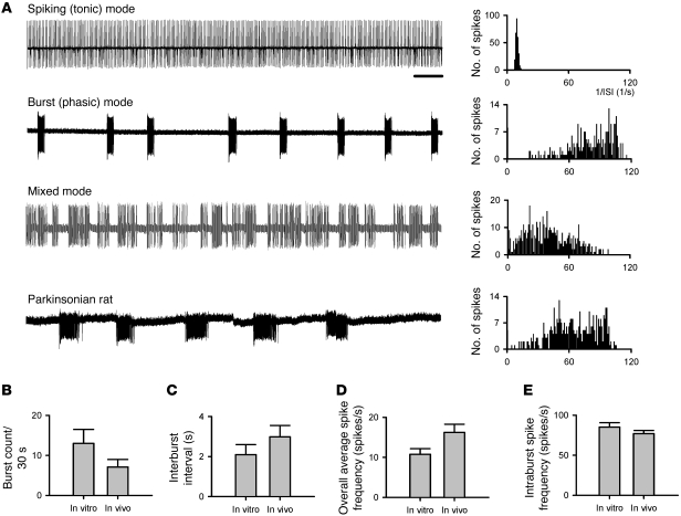
Figure 3. Spontaneous firing activities of subthalamic neurons in acute brain slices.
(A) Examples of cell-attached patch-clamp recordings of spontaneous subthalamic discharges in acute brain slices of normal mice and of parkinsonian rats (each sweep represents a 30-second recording). Subthalamic neurons in normal mice may exhibit spiking (tonic), burst (phasic), or mixed patterns of firing, whereas the neurons in parkinsonian rats tend to be either silent or firing in bursts with longer intra-burst duration. The corresponding histogram plot for each of the sample traces is presented on the right end of each trace. The histogram plot shows a clear peak in the inverse of the inter-spike interval (ISI) for the spiking (tonic) firing, representing regular spikes at a frequency of approximately 10 Hz. In contrast, there is a smear in the higher frequencies plus a single peak at an extremely low frequency for the burst firing mode, consistent with periodic occurrence of irregular but high-frequency bursts of discharges. Scale bar: 2 seconds. (B–E) Comparison of different parameters of STN firing recorded from acute slices (in vitro, n = 6) with those from in vivo single-unit recording data (taken from Figures 7 and 8). P = 0.24, 0.53, 0.31, and 0.41 for B, C, D, and E, respectively.

