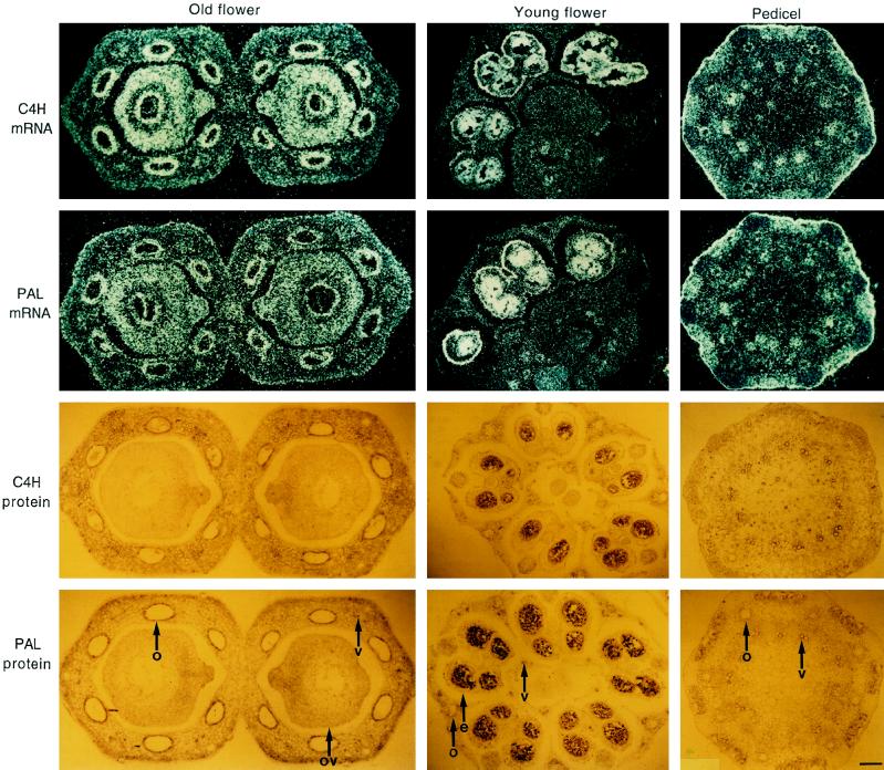
Figure 5.
In situ localization of C4H and PAL mRNA (top) and protein (bottom), respectively, in cross-sections from selected parsley tissues as indicated. 35S-Labeled antisense riboprobes and antisera were the same as described in Figures 3 and 4, respectively. e, Epidermis; o, oil-duct epithelial cells; ov, ovary; and v, vascular tissue. Magnification bar = 100 μm.

