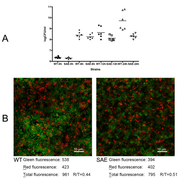
Figure 5.
Viability of S. epidermidis 1457 in biofilms and the planktonic state. (A) CFU counts of SE1457ΔsaeRS and SE1457. After 0, 6, 12, and 24 h of incubation, CFUs for SE1457 and SE1457ΔsaeRS cultures were calculated using serial dilutions of each sample plated on 6 agar plates. (B) CLSM images of S. epidermidis biofilms. SE1457 and SE1457ΔsaeRS were incubated in glass-bottomed cell culture dishes. After incubation at 37°C for 24 h, SE1457ΔsaeRS and SE1457 cells in biofilms were stained with LIVE/DEAD reagents that indicate viable cells by green fluorescence (SYTO9) and dead cells by red fluorescence (PI). Results depict a stack of images taken at approximately 0.3 μm depth increments and represent one of the three experiments. Fluorescence intensities were quantified using ImageJ software. WT, SE1457; SAE, SE1457ΔsaeRS.

