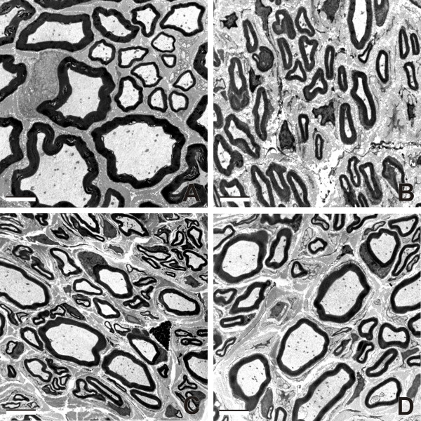
Figure 3.
Representative ultrathin sections through MCN. Electron micrographs showing representative myelinated axons in cross sections through intact MCN (A), and MCN stumps 2 months after their reconnection with the UN and intrathecal application of vehiculum (B), Cerebrolysin (C) and CNTF (D). Scale bars = 2 μm.

