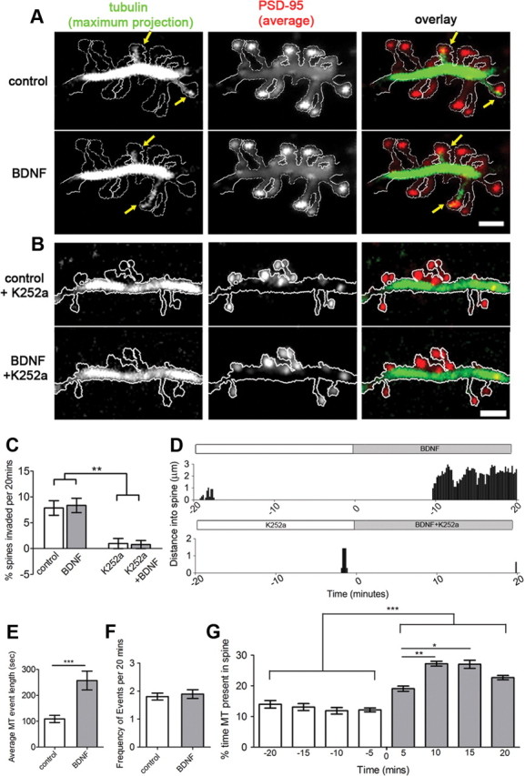
Figure 2.

Live-cell TIRFM shows increased MT invasions into spines treated with BDNF. A, A segment of dendrite from a 16 DIV hippocampal neuron, transfected with mCherry-α-tubulin and GFP-PSD-95, was imaged for 20 min before (control) and 20 min after addition of BDNF. Maximum projection of tubulin fluorescence depicts spines that were invaded by MTs (yellow arrows) during the time lapse. Average projection of PSD-95 fluorescence acts as a volume fill. B, A dendritic segment of a different neuron incubated with the tyrosine kinase inhibitor K252a for 24 h before the experiment. C, BDNF treatment does not change the percentage of spines invaded by MTs, whereas K252a decreases MT invasions (two-way ANOVA test with Bonfferoni post-tests). D, Examples of MT invasions into two dendritic spines. E, F, Average MT invasion event length is significantly increased after BDNF treatment (E), while the frequency of invasions among MT-targeted spines remains the same (F) (Student's t test). G, MT dwell time is elevated within 5 min of BDNF treatment and peaks at 10–15 min (Kruskal–Wallis test with Dunn's post hoc tests). Scale bars, 2 μm.
