
Fig. 4.
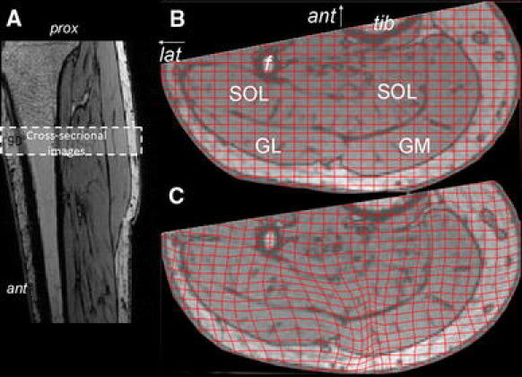
Typical example of MRI images with superimposed undeformed and deformed grids. a Longitudinal image of the lower leg, indicating the location of the cross-sectional images taken. b Cross-sectional MRI image of the lower leg (most proximal image of the stack) in the undeformed state, with the undeformed grid imposed and indications of anatomical structures to be seen. Knee joint angle ≈173 Ankle joint angle = 90°. c Cross-sectional MRI image of the lower leg (most proximal image of the stack) in the deformed state, with the deformed grid imposed (Knee joint angle ≈ 150°. Ankle joint angle = 90°). “Ant” indicates anterior side or direction (arrow) and “lat” indicates lateral side and direction (arrow “Tib” and “f” indicate tibia and fibula, respectively, and the relevant muscles are indicated as follows: GL and GM represent m. gastrocnemius caput lateralis and medialis, respectively, and SOL indicates m. soleus