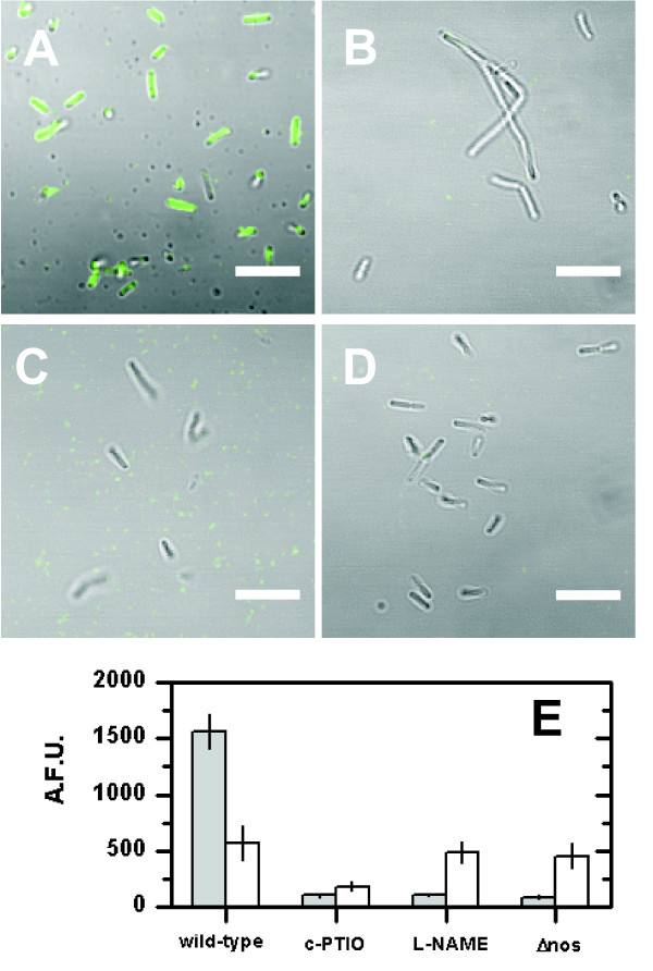
Figure 1.
Nitric-oxide-synthase (NOS)- derived NO formation by B. subtilis 3610. (A-D) Confocal laser scanning micrographs of cells grown in LB for 4 h at 37°C. Shown is the overlay of: gray - transmission and green - fluorescence of NO sensitive dye CuFL. (A) Wild-type without supplements, (B) supplemented with 100 μM c-PTIO (NO scavenger), (C) 100 μM L-NAME (NOS inhibitor), and (D) 3610Δnos. Scale bar is 5 μm. (E) Single-cell quantification of intracellular NO formation of cells grown in LB (gray bars) and MSgg (white bars) using CuFL fluorescence intensity (A.F.U. = Arbitrary Fluorescence Units). Error bars show standard error (N = 5).

