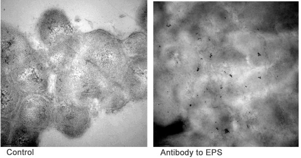
Figure 8.
Immuno-transmission electron micrographs of the OCT cryosection of an H. somni biofilm.H. somni was grown as a biofilm on glass slides and embedded in OCT resin to maintain the integrity of the biofilm prior to incubation with antiserum. Left, control OCT cryosection of biofilm incubated without specific antiserum, but with anti-rabbit conjugated gold particles; no labeling with the gold particles occurred; Right, OCT cryosection of a biofilm incubated with rabbit antibodies to EPS, followed by anti-rabbit conjugated gold particles. The black dots are gold particles around the bacterial cells and in the residual biofilm matrix.

