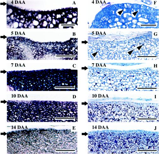
Figure 4.
In situ localization of the RINO1 transcript detected by a DIG-labeled antisense probe (A–E) and globoids detected by toluidine blue staining (F–J) in the scutellum of embryos 4 (A and F), 5 (B and G), 7 (C and H), 10 (D and I), and 14 DAA (E and J). Arrows indicate the border between the scutellum (bottom) and epithelium (top). Some of the globoids in F and G are marked with arrowheads. Scale bars represent 10 μm for A and F and 50 μm for B to E and G to J.

