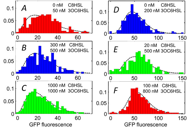
Figure 6.
Cell-to-cell heterogeneity at fixed average activation. Cell fluorescence brightness distributions measured for experiments #2 and #3 in Figure 4B. (A)-(C) Distributions collected for two-HSL combinations that generated ~25% of full lux activation (Experiment #2) and (D)-(F) distributions collected for combinations that generated ~60% of full lux activation (Experiment #3). The dashed curves show maximum likelihood fits to a gamma distribution. Each histogram is derived from roughly 200 individual cells.

