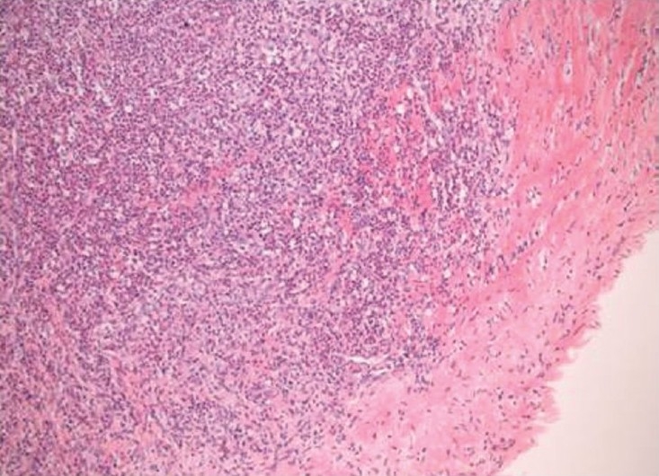
Figure 3.

Haematoxylin and eosin stain (×100) showing a dense inflammatory cell infiltrate including acute and chronic inflammatory cells with histiocytes and focal giant cells involving the vascular intima and extending into the rim of media included in the specimen.
