Figure 3.

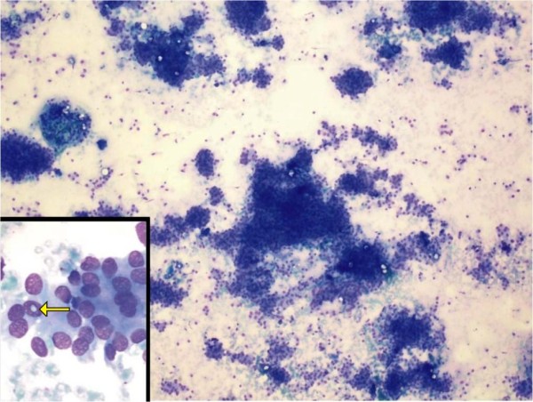
Photomicrograph showing moderately cellular smears composed of cells arranged in loose clusters, sheets and occasional whorls (hematoxylin and eosin stain; original magnification, ×100). Inset shows intra-nuclear inclusion (arrow).

Photomicrograph showing moderately cellular smears composed of cells arranged in loose clusters, sheets and occasional whorls (hematoxylin and eosin stain; original magnification, ×100). Inset shows intra-nuclear inclusion (arrow).