
Figure 3.
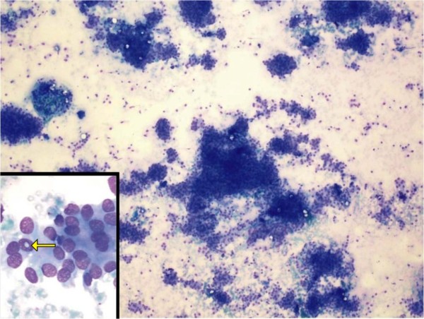
Photomicrograph showing moderately cellular smears composed of cells arranged in loose clusters, sheets and occasional whorls (hematoxylin and eosin stain; original magnification, ×100). Inset shows intra-nuclear inclusion (arrow).
Official websites use .gov
A
.gov website belongs to an official
government organization in the United States.
Secure .gov websites use HTTPS
A lock (
) or https:// means you've safely
connected to the .gov website. Share sensitive
information only on official, secure websites.
Photomicrograph showing moderately cellular smears composed of cells arranged in loose clusters, sheets and occasional whorls (hematoxylin and eosin stain; original magnification, ×100). Inset shows intra-nuclear inclusion (arrow).