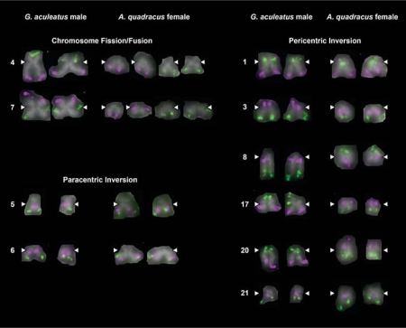
Fig. 2.
Major chromosomal rearrangements between G. aculeatus and A. quadracus. Two-color FISH images are shown for the 10 G. aculeatus chromosomes that show visible evidence for chromosome rearrangement. Pairs of homologs from individual metaphase spreads of G. aculeatus males and A. quadracus females are shown. White arrowheads indicate the position of the centromere.

