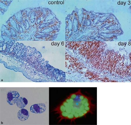
Fig. 1.
Location and morphology of mΦ in healthy colon. a F4/80+ cells in control mouse colon and 3, 6 and 8 days after induction of DSS colitis, showing the presence of mΦ in healthy intestine and the intense infiltrate in inflammation. Reproduced with permission from Stevceva et al. [109]. b Purified F4/80+ class II MHC+ mΦ from normal mouse colon show the morphological appearances of activation, with abundant foamy cytoplasmic granules and they phagcoytose FITC-labelled zymosan particles.

