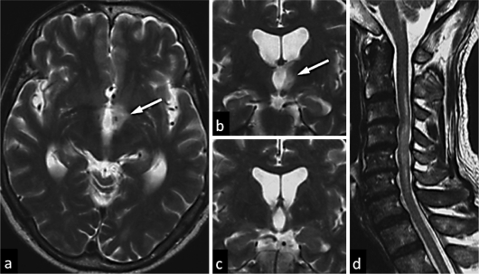
Fig. 1.
MRI findings of the present case. A hypothalamic lesion (arrow) was observed during the SIADH episode. Axial and coronal sections of a T2-weighted image showed marked hyperintense lesions at the hypothalamus (a, b). The hypothalamic lesion was markedly diminished during a relapse of acute myelitis 3 months after the SIADH episode (c). A sagittal T2-weighted image showed a long spinal cord lesion with high signal intensity during a relapse of acute myelitis (d).

