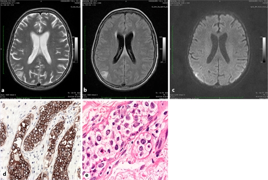
Fig. 2.
a–c Case 2. T2-weighted (a), fluid-attenuated inversion recovery (b) and diffusion-weighted (c) images showing a triangular shaped cortically based lesion with moderately restricted diffusion in the right parietal lobe suggestive of a subacute ischemic lesion. d Splenectomy specimen: blastic lymphoid tumor cells strictly confined to hilar blood vessels showing strong immunoreactivity with the pan-B cell marker CD20. e Skin biopsy: IVBCL infiltration in the deeper dermis.

