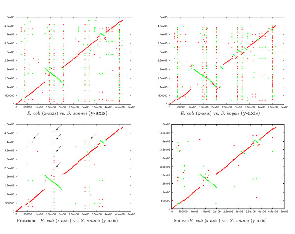
Figure 4.
Comparison of three bacterial genomes. A comparison of the three bacterial genomes of E. coli, S. sonnei, and S. boydii. We show the projections E. coli vs. S. sonnei (upper left) and E. coli vs. S. boydii (upper right). (The third projection S. sonnei vs. S. boydii is not shown). The plot on the lower-left is the projection E. coli vs. S. sonnei, where each point represents a protein that is encoded in all three genomes. (Other projections are not shown.). The arrows point to some proteins repeated in the genomes (vertically aligned hits correspond to repetitions in S. sonnei and the horizontally aligned ones correspond to repetitions in E. coli.). The fourth plot is the projection E. coli vs. S. sonnei plotted according to the alignment computed by the program Mauve. Red lines correspond to similar regions (or protein hits) between the forward strands of the genomes on the x-and y-axis, while green lines correspond to similar regions (or protein hits) between the forward strand of the genomes on the x-axis and the backward strand on the y-axis.

