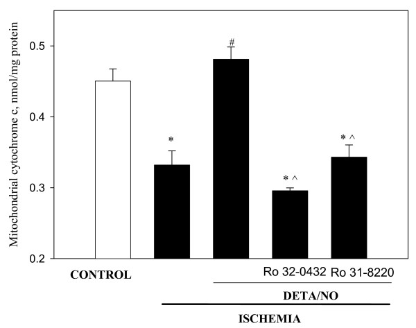
Figure 5.
Perfusion of the hearts with PKC inhibitors abolish the protective effect of DETA/NO against ischemia-induced loss of cytochrome c from mitochondria. Hearts were perfused 15 min with 0.5 μM Ro 32-0432 and 1 μM Ro 31-8220 followed by 3 min perfusion with 50 μM DETA/NO. Then hearts were subjected to 30 min stop-flow ischemia. Mitochondrial content of cytochrome c was measured as described in Methods. * – Statistically significant effect of ischemia if compared to control (p < 0.01, Tukey test); # – statistically significant effect of DETA/NO if compared to ischemia (p < 0.01, Tukey test); ^ – statistically significant effect of PKC inhibitors if compared to DETA/NO group (p < 0.05, Tukey test). Means ± standard errors of 4 separate experiments are presented.

